Microscopy Beyond the Monochromatic Lens: A Kaleidoscopic Experience

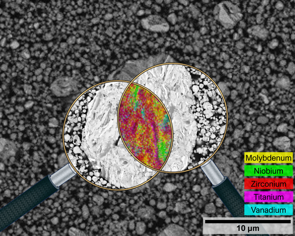
This is a cross-sectional micrograph of a metal alloy particle with a size nearly equal to that of a white blood cell (approx. 12 microns). Showcasing the power of a scanning electron microscope, the developed metal alloy particles are observed at very high magnifications to confirm the alloying or incorporation of different elements within the particle. The metal alloy particles are formed from a continuous cycle of fracturing and welding of elemental particles in which the microstructures can be seen in the micrograph. Interestingly, beyond the regular monochromatic micrographs, a scanning electron microscope also has the capability to provide a kaleidoscopic micrograph showing the elemental composition of the metal alloy particle in terms of a colorful spectrum. The data obtained from the scanning electron microscope serve as important information in the research and development of metal alloy powders for various industrial applications.